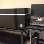
The DRSC/TRiP-FGR is pleased to announce that we recently updated our high-throughput, high-content imaging system. Through funds from NIH, HHMI, and the Harvard Medical School Tools & Technology program, we were able to get a GE IN Cell 6000 automated imaging system.
The instrument in the configuration at the DRSC includes a PAA KiNEDx robotic plate handler and 4x, 10x, 20x, and 60x lenses. The instrument lets users mix-and-match among epifluorescence, confocal fluorescence, and bright field imaging. The instrument supports imaging from microscope slides (up to four at a time) and from micro-well plates with wells of many sizes, from 6- and 12-well on up to 96- and 384-well formats.
This powerful system is already supporting a next generation of high-content image-based screens and smaller projects. Because it allows for imaging with microscope slides as well as micro-well plates, the system is appropriate for both in vivo and cultured or primary cell-based studies. Access is open to all, including local researchers and visiting screeners.
