Cell-based assays

Regardless of the technology (RNAi, CRISPR, over-expression, etc.), a good cell-based assay is the best foundation for a cell-based screen. We have equipment, provide reagents, share protocols, and more to support development of high-throughput screen assays in Drosophila cells.

Reagents, consultation, and other support is available for screens off-site. We also support screens on-site at our facility. Assays can be done using a number of types of reagents, including reagents for knockdown or over-expression of protein-coding genes, and interrogation of miRNAs.

See links below to relevant reagents, protocols, publications, and more.

News

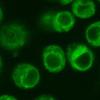
Image of green fluorescence in GFP-tagged Drosophila cultured cells

New cell lines & new understandings using cutting-edge techniques

February 2, 2024

As a facility that supports large-scale screens in Drosophila and other insect cell lines, we get excited about reports of new Drosophila cell lines and related info.

We'd like to highlight two recent papers.

One report, a collaboration between Amanda Simcox's group, the DGRC, and our group here at the DRSC, describes new cell lines made in Amanda's group and characterized in a collaboration of the three groups. Muscle cells that pulse? Yes. That and other exciting new cell lines are reported in the publication below, and the cells are available at the DGRC...

Read more about New cell lines & new understandings using cutting-edge techniques
Decorative cartoon drawn with BioRender depicting DRSC-BTRR technology concepts

So you want to do a CRISPR pooled screen in insect cells? You can! Here's how

May 12, 2022

At the DRSC-BTRR, we've been doing a lot of pooled-format CRISPR knockout screens in Drosophila cells. We're finding the results to be robust and reproducible. And best of all, the results have been informative, providing insights into diverse areas of biology.

Thinking about how to do CRISPR knockout screens in cells is a little different from thinking about how to do a genetic or RNAi screen in vivo or doing an arrayed-format RNAi screen....

Read more about So you want to do a CRISPR pooled screen in insect cells? You can! Here's how
Graphical image of tissue culture, fly pushing, and computer, and the team of people who work with them

DRSC/TRiP and DRSC-BTRR Office Hours

September 13, 2021

New this fall: Online office hours!

Do you have questions about modifying Drosophila cell lines with CRISPR or performing large-scale cell screens? Questions about in vivo RNAi with TRiP fly stocks or CRISPR knockout or activation with our sgRNA fly stocks? Questions about our new protocols and resources for CRISPR mosquito cell lines? Pop into our Zoom office hours to say hello and get our expert input! Registration is required (see below).

DRSC/TRiP & DRSC-BTRR Office Hours Schedule:

Mon. Sept. 27, 2021, 12...

Read more about DRSC/TRiP and DRSC-BTRR Office Hours
Graphical image of tissue culture, fly pushing, and computer, and the team of people who work with them

DRSC-Biomedical Technology Research Resource

October 21, 2019

We are pleased to announce that we have been funded by NIH NIGMS to form the Drosophila Research & Screening Center-Biomedical Technology Research Resource (DRSC-BTRR). The P41-funded DRSC-BTRR (N. Perrimon, PI; S. Mohr, Co-I) builds upon and extends past goals of the Drosophila RNAi Screening Center.

As the DRSC-BTRR, we are working together with collaborators whose 'driving biomedical projects' inform development of new technologies at the DRSC. At the same time, we continue to support Drosophila cell-based RNAi and CRIPSR knockout screens and related...

Read more about DRSC-Biomedical Technology Research Resource
Photo of 384-well assay plates

Drosophila cell screen with DRSC reagent library contributes to identification of new therapeutic target for renal cancer

October 7, 2019

We here at the DRSC/TRiP are thrilled to see this study from Hilary Nicholson et al. published in Science Signaling.

The study provides a great example of how screens in Drosophila cultured cells can be used as part of a cross-species platform aimed at discovery of new targets for disease treatment. The work represents a collaboration between the laboratory of 2019 Nobel Prize winner W. Kaelin and DRSC PI N. Perrimon.

...

Read more about Drosophila cell screen with DRSC reagent library contributes to identification of new therapeutic target for renal cancer

Contact Us

Please contact us for any questions.

Publications

Agustin Rolandelli, Hanna J Laukaitis-Yousey, Haikel N Bogale, Nisha Singh, Sourabh Samaddar, Anya J O’Neal, Camila R Ferraz, Matthew Butnaru, Enzo Mameli, Baolong Xia, Tays M. Mendes, Rainer L. Butler, Liron Marnin, Francy ECabrera Paz, Luisa M Valencia, Vipin S Rana, Ciaran Skerry, Utpal Pal, Stephanie E Mohr, Norbert Perrimon, David Serre, and Joao HF Pedra. 2023. “Tick hemocytes have pleiotropic roles in microbial infection and arthropod fitness.” bioRxiv, Pp. 2023.08.31.555785. Publisher's VersionAbstract
Uncovering the complexity of systems in non-model organisms is critical for understanding arthropod immunology. Prior efforts have mostly focused on Dipteran insects, which only account for a subset of existing arthropod species in nature. Here, we describe immune cells or hemocytes from the clinically relevant tick Ixodes scapularis using bulk and single cell RNA sequencing combined with depletion via clodronate liposomes, RNA interference, Clustered Regularly Interspaced Short Palindromic Repeats activation (CRISPRa) and RNA-fluorescence in situ hybridization (FISH). We observe molecular alterations in hemocytes upon tick infestation of mammals and infection with either the Lyme disease spirochete Borrelia burgdorferi or the rickettsial agent Anaplasma phagocytophilum. We predict distinct hemocyte lineages and reveal clusters exhibiting defined signatures for immunity, metabolism, and proliferation during hematophagy. Furthermore, we perform a mechanistic characterization of two I. scapularis hemocyte markers: hemocytin and astakine. Depletion of phagocytic hemocytes affects hemocytin and astakine levels, which impacts blood feeding and molting behavior of ticks. Hemocytin specifically affects the c-Jun N-terminal kinase (JNK) signaling pathway, whereas astakine alters hemocyte proliferation in I. scapularis. Altogether, we uncover the heterogeneity and pleiotropic roles of hemocytes in ticks and provide a valuable resource for comparative biology in arthropods.Competing Interest StatementThe authors have declared no competing interest.
Nikki Coleman-Gosser, Yanhui Hu, Shiva Raghuvanshi, Shane Stitzinger, Weihang Chen, Arthur Luhur, Daniel Mariyappa, Molly Josifov, Andrew Zelhof, Stephanie E Mohr, Norbert Perrimon, and Amanda Simcox. 2023. “Continuous muscle, glial, epithelial, neuronal, and hemocyte cell lines for research.” Elife, 12.Abstract

Expression of activated Ras, Ras, provides cultured cells with a proliferation and survival advantage that simplifies the generation of continuous cell lines. Here, we used lineage-restricted Ras expression to generate continuous cell lines of muscle, glial, and epithelial cell type. Additionally, cell lines with neuronal and hemocyte characteristics were isolated by cloning from cell cultures established with broad Ras expression. Differentiation with the hormone ecdysone caused maturation of cells from mesoderm lines into active muscle tissue and enhanced dendritic features in neuronal-like lines. Transcriptome analysis showed expression of key cell-type-specific genes and the expected alignment with single-cell sequencing and in situ data. Overall, the technique has produced in vitro cell models with characteristics of glia, epithelium, muscle, nerve, and hemocyte. The cells and associated data are available from the Genomic Resource Center.

Baolong Xia, Raghuvir Viswanatha, Yanhui Hu, Stephanie E Mohr, and Norbert Perrimon. 2023. “Pooled genome-wide CRISPR activation screening for rapamycin resistance genes in cells.” Elife, 12.Abstract

Loss-of-function and gain-of-function genetic perturbations provide valuable insights into gene function. In cells, while genome-wide loss-of-function screens have been extensively used to reveal mechanisms of a variety of biological processes, approaches for performing genome-wide gain-of-function screens are still lacking. Here, we describe a pooled CRISPR activation (CRISPRa) screening platform in cells and apply this method to both focused and genome-wide screens to identify rapamycin resistance genes. The screens identified three genes as novel rapamycin resistance genes: a member of the SLC16 family of monocarboxylate transporters (), a member of the lipocalin protein family (), and a zinc finger C2H2 transcription factor (). Mechanistically, we demonstrate that overexpression activates the RTK-Akt-mTOR signaling pathway and that activation of insulin receptor (InR) by requires cholesterol and clathrin-coated pits at the cell membrane. This study establishes a novel platform for functional genetic studies in cells.

Shue Chen, Leah F Rosin, Gianluca Pegoraro, Nellie Moshkovich, Patrick J Murphy, Guoyun Yu, and Elissa P Lei. 8/12/2022. “NURF301 contributes to gypsy chromatin insulator-mediated nuclear organization.” Nucleic Acids Res, 50, 14, Pp. 7906-7924.Abstract
Chromatin insulators are DNA-protein complexes that can prevent the spread of repressive chromatin and block communication between enhancers and promoters to regulate gene expression. In Drosophila, the gypsy chromatin insulator complex consists of three core proteins: CP190, Su(Hw), and Mod(mdg4)67.2. These factors concentrate at nuclear foci termed insulator bodies, and changes in insulator body localization have been observed in mutants defective for insulator function. Here, we identified NURF301/E(bx), a nucleosome remodeling factor, as a novel regulator of gypsy insulator body localization through a high-throughput RNAi imaging screen. NURF301 promotes gypsy-dependent insulator barrier activity and physically interacts with gypsy insulator proteins. Using ChIP-seq, we found that NURF301 co-localizes with insulator proteins genome-wide, and NURF301 promotes chromatin association of Su(Hw) and CP190 at gypsy insulator binding sites. These effects correlate with NURF301-dependent nucleosome repositioning. At the same time, CP190 and Su(Hw) both facilitate recruitment of NURF301 to chromatin. Finally, Oligopaint FISH combined with immunofluorescence revealed that NURF301 promotes 3D contact between insulator bodies and gypsy insulator DNA binding sites, and NURF301 is required for proper nuclear positioning of gypsy binding sites. Our data provide new insights into how a nucleosome remodeling factor and insulator proteins cooperatively contribute to nuclear organization.
Ying Xu, Raghuvir Viswanatha, Oleg Sitsel, Daniel Roderer, Haifang Zhao, Christopher Ashwood, Cecilia Voelcker, Songhai Tian, Stefan Raunser, Norbert Perrimon, and Min Dong. 2022. “CRISPR screens in Drosophila cells identify Vsg as a Tc toxin receptor.” Nature, 610, 7931, Pp. 349-355.Abstract
Entomopathogenic nematodes are widely used as biopesticides1,2. Their insecticidal activity depends on symbiotic bacteria such as Photorhabdus luminescens, which produces toxin complex (Tc) toxins as major virulence factors3-6. No protein receptors are known for any Tc toxins, which limits our understanding of their specificity and pathogenesis. Here we use genome-wide CRISPR-Cas9-mediated knockout screening in Drosophila melanogaster S2R+ cells and identify Visgun (Vsg) as a receptor for an archetypal P. luminescens Tc toxin (pTc). The toxin recognizes the extracellular O-glycosylated mucin-like domain of Vsg that contains high-density repeats of proline, threonine and serine (HD-PTS). Vsg orthologues in mosquitoes and beetles contain HD-PTS and can function as pTc receptors, whereas orthologues without HD-PTS, such as moth and human versions, are not pTc receptors. Vsg is expressed in immune cells, including haemocytes and fat body cells. Haemocytes from Vsg knockout Drosophila are resistant to pTc and maintain phagocytosis in the presence of pTc, and their sensitivity to pTc is restored through the transgenic expression of mosquito Vsg. Last, Vsg knockout Drosophila show reduced bacterial loads and lethality from P. luminescens infection. Our findings identify a proteinaceous Tc toxin receptor, reveal how Tc toxins contribute to P. luminescens pathogenesis, and establish a genome-wide CRISPR screening approach for investigating insecticidal toxins and pathogens.
Hans M Dalton, Raghuvir Viswanatha, Roderick Brathwaite, Jae Sophia Zuno, Alexys R Berman, Rebekah Rushforth, Stephanie E Mohr, Norbert Perrimon, and Clement Y Chow. 2022. “A genome-wide CRISPR screen identifies DPM1 as a modifier of DPAGT1 deficiency and ER stress.” PLoS Genet, 18, 9, Pp. e1010430.Abstract
Partial loss-of-function mutations in glycosylation pathways underlie a set of rare diseases called Congenital Disorders of Glycosylation (CDGs). In particular, DPAGT1-CDG is caused by mutations in the gene encoding the first step in N-glycosylation, DPAGT1, and this disorder currently lacks effective therapies. To identify potential therapeutic targets for DPAGT1-CDG, we performed CRISPR knockout screens in Drosophila cells for genes associated with better survival and glycoprotein levels under DPAGT1 inhibition. We identified hundreds of candidate genes that may be of therapeutic benefit. Intriguingly, inhibition of the mannosyltransferase Dpm1, or its downstream glycosylation pathways, could rescue two in vivo models of DPAGT1 inhibition and ER stress, even though impairment of these pathways alone usually causes CDGs. While both in vivo models ostensibly cause cellular stress (through DPAGT1 inhibition or a misfolded protein), we found a novel difference in fructose metabolism that may indicate glycolysis as a modulator of DPAGT1-CDG. Our results provide new therapeutic targets for DPAGT1-CDG, include the unique finding of Dpm1-related pathways rescuing DPAGT1 inhibition, and reveal a novel interaction between fructose metabolism and ER stress.
  •  
  • 1 of 19
  • »
More