*UPDATE* The IN Cell Analyzer remains available to the research community and in our lab space. However, it is now being managed by the HMS Microscopy in the North Quad (MicRoN) Core facility. Visit the MicRoN core facility website for contact info and other details.
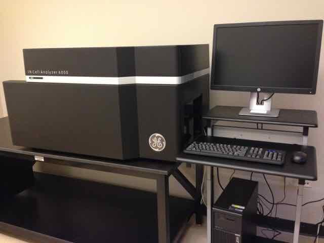
High-content, high-throughput imaging at the DRSC/TRiP-FGR is supported by a GE IN Cell Analyzer 6000 Cell Imaging System with automation [UPDATE: access by image-based screeners to the IN Cell is now managed by the MicRoN core]. The IN Cell has 4x, 10x, 20x, and 60x lenses, and supports multi-color confocal or non-confocal fluorescence imaging as well as brightfield imaging. Please use the order/signup system or get in touch with the Director to learn about using this instrument for low- or high-throughput screens and other imaging projects.

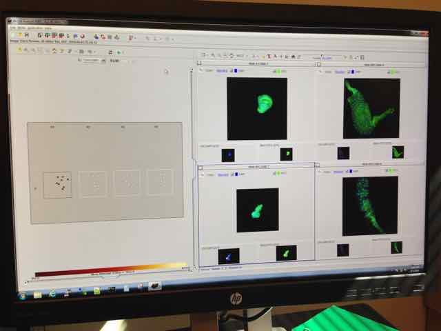
In addition to supporting confocal imaging of cells in 96-and 384-well plates, the IN Cell 6000 also supports imaging of standard microscope slides, 6-well dishes, and other lower throughput platforms. Shown here, imaging at 10x of a microscope slide with GFP and DAPI in larval tissues. To the left, you can see a map of the coverslip, with the fields of view indicated. To the right, quick view of images from the slide. Visit the MicRoN core facility website for contact info and other details.

