Image analysis

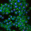
Figure 1 from Xu, Kim et al 2022 in eLife

Light up your flies! Reagent and protocol information for application of our NanoTag system for epitope tagging in Drosophila

March 23, 2023

One of the areas of interest for our technology development group is nanobodies. These small, single-chain antibodies are particularly attractive for Drosophila research as in addition to being used for standard immune-technologies such as immunoblots and immunostaining of tissues, they can also be expressed in vivo as fusions to fluorescent proteins (‘chromobodies’) or functional domains (e.g., for degradation or re-localization).

Past applications of this technology in Drosophila relied on availability of nanobodies targeting a specific protein or use of nanobodies targeting...

Read more about Light up your flies! Reagent and protocol information for application of our NanoTag system for epitope tagging in Drosophila
2018 Dec 08

DRSC&TRiP at ASCB|EMBO 2018

Sat Dec 8 (All day) to Wed Dec 12 (All day)

Location: 

San Diego, CA, USA
The DRSC & TRiP will be represented at the ASCB|EMBO 2018 conference in the form of a poster that includes information about our collaboration with O. Kanca in the H. Bellen lab at Baylor College of Medicine to tag Drosophila S2R+ cell lines with GFP using a CRISPR-based strategy. We have 'painted' a number of subcellular localizations green with this approach! If you're at the meeting, check out P1482 at Board Number B494, presented by Baolong Xia of the Perrimon lab.
Joel M Swenson, Serafin U Colmenares, Amy R Strom, Sylvain V Costes, and Gary H Karpen. 2016. “The composition and organization of Drosophila heterochromatin are heterogeneous and dynamic.” Elife, 5.Abstract

Heterochromatin is enriched for specific epigenetic factors including Heterochromatin Protein 1a (HP1a), and is essential for many organismal functions. To elucidate heterochromatin organization and regulation, we purified Drosophila melanogaster HP1a interactors, and performed a genome-wide RNAi screen to identify genes that impact HP1a levels or localization. The majority of the over four hundred putative HP1a interactors and regulators identified were previously unknown. We found that 13 of 16 tested candidates (83%) are required for gene silencing, providing a substantial increase in the number of identified components that impact heterochromatin properties. Surprisingly, image analysis revealed that although some HP1a interactors and regulators are broadly distributed within the heterochromatin domain, most localize to discrete subdomains that display dynamic localization patterns during the cell cycle. We conclude that heterochromatin composition and architecture is more spatially complex and dynamic than previously suggested, and propose that a network of subdomains regulates diverse heterochromatin functions.

Mar Arias Garcia, Miguel Sanchez Alvarez, Heba Sailem, Vicky Bousgouni, Julia Sero, and Chris Bakal. 2012. “Differential RNAi screening provides insights into the rewiring of signalling networks during oxidative stress.” Mol Biosyst, 8, 10, Pp. 2605-13.Abstract

Reactive Oxygen Species (ROS) are a natural by-product of cellular growth and proliferation, and are required for fundamental processes such as protein-folding and signal transduction. However, ROS accumulation, and the onset of oxidative stress, can negatively impact cellular and genomic integrity. Signalling networks have evolved to respond to oxidative stress by engaging diverse enzymatic and non-enzymatic antioxidant mechanisms to restore redox homeostasis. The architecture of oxidative stress response networks during periods of normal growth, and how increased ROS levels dynamically reconfigure these networks are largely unknown. In order to gain insight into the structure of signalling networks that promote redox homeostasis we first performed genome-scale RNAi screens to identify novel suppressors of superoxide accumulation. We then infer relationships between redox regulators by hierarchical clustering of phenotypic signatures describing how gene inhibition affects superoxide levels, cellular viability, and morphology across different genetic backgrounds. Genes that cluster together are likely to act in the same signalling pathway/complex and thus make "functional interactions". Moreover we also calculate differential phenotypic signatures describing the difference in cellular phenotypes following RNAi between untreated cells and cells submitted to oxidative stress. Using both phenotypic signatures and differential signatures we construct a network model of functional interactions that occur between components of the redox homeostasis network, and how such interactions become rewired in the presence of oxidative stress. This network model predicts a functional interaction between the transcription factor Jun and the IRE1 kinase, which we validate in an orthogonal assay. We thus demonstrate the ability of systems-biology approaches to identify novel signalling events.

Jianwu Bai, Richard Binari, Jian-Quan Ni, Marina Vijayakanthan, Hong-Sheng Li, and Norbert Perrimon. 2008. “RNA interference screening in Drosophila primary cells for genes involved in muscle assembly and maintenance.” Development, 135, 8, Pp. 1439-49.Abstract

To facilitate the genetic analysis of muscle assembly and maintenance, we have developed a method for efficient RNA interference (RNAi) in Drosophila primary cells using double-stranded RNAs (dsRNAs). First, using molecular markers, we confirm and extend the observation that myogenesis in primary cultures derived from Drosophila embryonic cells follows the same developmental course as that seen in vivo. Second, we apply this approach to analyze 28 Drosophila homologs of human muscle disease genes and find that 19 of them, when disrupted, lead to abnormal muscle phenotypes in primary culture. Third, from an RNAi screen of 1140 genes chosen at random, we identify 49 involved in late muscle differentiation. We validate our approach with the in vivo analyses of three genes. We find that Fermitin 1 and Fermitin 2, which are involved in integrin-containing adhesion structures, act in a partially redundant manner to maintain muscle integrity. In addition, we characterize CG2165, which encodes a plasma membrane Ca2+-ATPase, and show that it plays an important role in maintaining muscle integrity. Finally, we discuss how Drosophila primary cells can be manipulated to develop cell-based assays to model human diseases for RNAi and small-molecule screens.

Zheng Yin, Amine Sadok, Heba Sailem, Afshan McCarthy, Xiaofeng Xia, Fuhai Li, Mar Arias Garcia, Louise Evans, Alexis R Barr, Norbert Perrimon, Christopher J Marshall, Stephen TC Wong, and Chris Bakal. 2013. “A screen for morphological complexity identifies regulators of switch-like transitions between discrete cell shapes.” Nat Cell Biol, 15, 7, Pp. 860-71.Abstract

The way in which cells adopt different morphologies is not fully understood. Cell shape could be a continuous variable or restricted to a set of discrete forms. We developed quantitative methods to describe cell shape and show that Drosophila haemocytes in culture are a heterogeneous mixture of five discrete morphologies. In an RNAi screen of genes affecting the morphological complexity of heterogeneous cell populations, we found that most genes regulate the transition between discrete shapes rather than generating new morphologies. In particular, we identified a subset of genes, including the tumour suppressor PTEN, that decrease the heterogeneity of the population, leading to populations enriched in rounded or elongated forms. We show that these genes have a highly conserved function as regulators of cell shape in both mouse and human metastatic melanoma cells.

Mathias Beller, Carole Sztalryd, Noel Southall, Ming Bell, Herbert Jäckle, Douglas S Auld, and Brian Oliver. 2008. “COPI complex is a regulator of lipid homeostasis.” PLoS Biol, 6, 11, Pp. e292.Abstract

Lipid droplets are ubiquitous triglyceride and sterol ester storage organelles required for energy storage homeostasis and biosynthesis. Although little is known about lipid droplet formation and regulation, it is clear that members of the PAT (perilipin, adipocyte differentiation related protein, tail interacting protein of 47 kDa) protein family coat the droplet surface and mediate interactions with lipases that remobilize the stored lipids. We identified key Drosophila candidate genes for lipid droplet regulation by RNA interference (RNAi) screening with an image segmentation-based optical read-out system, and show that these regulatory functions are conserved in the mouse. Those include the vesicle-mediated Coat Protein Complex I (COPI) transport complex, which is required for limiting lipid storage. We found that COPI components regulate the PAT protein composition at the lipid droplet surface, and promote the association of adipocyte triglyceride lipase (ATGL) with the lipid droplet surface to mediate lipolysis. Two compounds known to inhibit COPI function, Exo1 and Brefeldin A, phenocopy COPI knockdowns. Furthermore, RNAi inhibition of ATGL and simultaneous drug treatment indicate that COPI and ATGL function in the same pathway. These data indicate that the COPI complex is an evolutionarily conserved regulator of lipid homeostasis, and highlight an interaction between vesicle transport systems and lipid droplets.

Inma Gonzalez, Julio Mateos-Langerak, Aubin Thomas, Thierry Cheutin, and Giacomo Cavalli. 2014. “Identification of regulators of the three-dimensional polycomb organization by a microscopy-based genome-wide RNAi screen.” Mol Cell, 54, 3, Pp. 485-99.Abstract

Polycomb group (PcG) proteins dynamically define cellular identities through epigenetic repression of key developmental genes. PcG target gene repression can be stabilized through the interaction in the nucleus at PcG foci. Here, we report the results of a high-resolution microscopy genome-wide RNAi screen that identifies 129 genes that regulate the nuclear organization of Pc foci. Candidate genes include PcG components and chromatin factors, as well as many protein-modifying enzymes, including components of the SUMOylation pathway. In the absence of SUMO, Pc foci coagulate into larger aggregates. Conversely, loss of function of the SUMO peptidase Velo disperses Pc foci. Moreover, SUMO and Velo colocalize with PcG proteins at PREs, and Pc SUMOylation affects its chromatin targeting, suggesting that the dynamic regulation of Pc SUMOylation regulates PcG-mediated silencing by modulating the kinetics of Pc binding to chromatin as well as its ability to form Polycomb foci.

Chaohong Wu, Joost Schulte, Katharine J Sepp, Troy J Littleton, and Pengyu Hong. 2010. “Automatic robust neurite detection and morphological analysis of neuronal cell cultures in high-content screening.” Neuroinformatics, 8, 2, Pp. 83-100.Abstract

Cell-based high content screening (HCS) is becoming an important and increasingly favored approach in therapeutic drug discovery and functional genomics. In HCS, changes in cellular morphology and biomarker distributions provide an information-rich profile of cellular responses to experimental treatments such as small molecules or gene knockdown probes. One obstacle that currently exists with such cell-based assays is the availability of image processing algorithms that are capable of reliably and automatically analyzing large HCS image sets. HCS images of primary neuronal cell cultures are particularly challenging to analyze due to complex cellular morphology. Here we present a robust method for quantifying and statistically analyzing the morphology of neuronal cells in HCS images. The major advantages of our method over existing software lie in its capability to correct non-uniform illumination using the contrast-limited adaptive histogram equalization method; segment neuromeres using Gabor-wavelet texture analysis; and detect faint neurites by a novel phase-based neurite extraction algorithm that is invariant to changes in illumination and contrast and can accurately localize neurites. Our method was successfully applied to analyze a large HCS image set generated in a morphology screen for polyglutamine-mediated neuronal toxicity using primary neuronal cell cultures derived from embryos of a Drosophila Huntington's Disease (HD) model.

Christophe J Echeverri and Norbert Perrimon. 2006. “High-throughput RNAi screening in cultured cells: a user's guide.” Nat Rev Genet, 7, 5, Pp. 373-84.Abstract

RNA interference has re-energized the field of functional genomics by enabling genome-scale loss-of-function screens in cultured cells. Looking back on the lessons that have been learned from the first wave of technology developments and applications in this exciting field, we provide both a user's guide for newcomers to the field and a detailed examination of some more complex issues, particularly concerning optimization and quality control, for more advanced users. From a discussion of cell lines, screening paradigms, reagent types and read-out methodologies, we explore in particular the complexities of designing optimal controls and normalization strategies for these challenging but extremely powerful studies.

Eric F Joyce, Benjamin R Williams, Tiao Xie, and C-Ting Wu. 2012. “Identification of genes that promote or antagonize somatic homolog pairing using a high-throughput FISH-based screen.” PLoS Genet, 8, 5, Pp. e1002667.Abstract

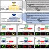
The pairing of homologous chromosomes is a fundamental feature of the meiotic cell. In addition, a number of species exhibit homolog pairing in nonmeiotic, somatic cells as well, with evidence for its impact on both gene regulation and double-strand break (DSB) repair. An extreme example of somatic pairing can be observed in Drosophila melanogaster, where homologous chromosomes remain aligned throughout most of development. However, our understanding of the mechanism of somatic homolog pairing remains unclear, as only a few genes have been implicated in this process. In this study, we introduce a novel high-throughput fluorescent in situ hybridization (FISH) technology that enabled us to conduct a genome-wide RNAi screen for factors involved in the robust somatic pairing observed in Drosophila. We identified both candidate "pairing promoting genes" and candidate "anti-pairing genes," providing evidence that pairing is a dynamic process that can be both enhanced and antagonized. Many of the genes found to be important for promoting pairing are highly enriched for functions associated with mitotic cell division, suggesting a genetic framework for a long-standing link between chromosome dynamics during mitosis and nuclear organization during interphase. In contrast, several of the candidate anti-pairing genes have known interphase functions associated with S-phase progression, DNA replication, and chromatin compaction, including several components of the condensin II complex. In combination with a variety of secondary assays, these results provide insights into the mechanism and dynamics of somatic pairing.

Pages